Eye Health and Diseases

Eye Health and Diseases

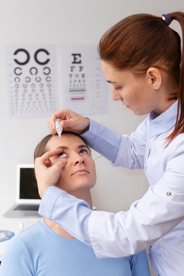
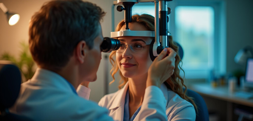
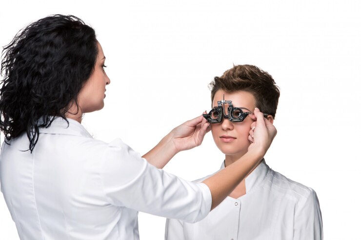
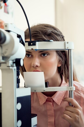
At the eye diseases department , the initial step in a routine eye examination involves discussing the patient's visual complaints. Based on the nature of these complaints, the examination begins with observations of the patient's eyebrows, eyelids, and eye positioning. The patient's refractive error is accurately measured using a computerized auto-refractometer and a retinoscope. Subsequently, visual acuity for each eye is assessed with and without corrective lenses. A detailed biomicroscopic examination is then conducted to evaluate the eyelashes, conjunctiva, cornea, and other components of the eye's anterior segment, followed by intraocular pressure measurement.

Refractive Errors

  • Light and images are refracted by the cornea and lens to focus on the retina, facilitating vision. In an ideal scenario, this process aligns precisely with the visual cortex to optimize visual acuity.
  • Refractive errors can impair the cornea, lens, retina, or optic nerve, necessitating regular comprehensive eye examinations.
  • Common symptoms include visual disturbances, eye pain, and discomfort, categorized into myopia (nearsightedness), hypermetropia (farsightedness), astigmatism, and presbyopia (age-related farsightedness).
  • Correction options include eyeglasses, contact lenses, and excimer laser treatments. These examinations also facilitate the diagnosis of conditions like retinal detachment, hypertension, and brain tumors.

Cataract and Its Treatment (Phacoemulsification)

  • A cataract is characterized by the clouding of the eyes normally clear lens, (predominantly due to aging, but can affect all ages, including infants). Symptoms include painless Visual loss (painless), glare, fading of color and poor nightly vision.
  • The definitive treatment is phacoemulsification surgery, a cutting-edge technique that involves the insertion of a high-quality, FDA-approved intraocular lens, potentially including multifocal or trifocal options, depending on patient preference.

Glaucoma and Its Management

  • Glaucoma results from inadequate drainage of intraocular fluid, leading to increased pressure and optic nerve damage, potentially causing irreversible blindness. Symptoms may include blurred vision, severe eye pain, headaches, nausea, and vomiting.
  • Regular eye check-ups are crucial for early detection. Treatment options to prevent further vision loss include medicated eye drops, argon laser surgery, and other surgical procedures. Diagnostic tools include Optical Coh.

Pediatric Ophthalmology

  • Specialized pediatric autorefractometers provide an effective means for assessing children's eyesight, with essential routine screenings recommended at birth, and at ages one and three. These screenings are vital for diagnosing and treating vision issues, particularly in children with a family history of eye problems.

Strabismus

  • Strabismus involves misalignment of the eyes, which may result from not wearing needed glasses, muscle anomalies, or congenital/neurological issues. Treatment aims to improve vision, correct head posture, eliminate double vision, enable proper eye movement, and address aesthetic concerns through surgical methods.

Oculoplastic Surgery

  • This field covers treatments for eyelid malpositions, wrinkle correction, under-eye bag removal, lacrimal duct obstructions, eyelid malformations, ocular prostheses, and eye tumor treatments.

Retinal Diseases

  • The retina is akin to a camera sensor, capturing light and sending images to the brain via the optic nerve, with the macula enabling central, sharp vision. The vitreous, a gel-like substance, fills the eye and supports its shape.
  • Common retinal conditions include diabetic retinopathy, retinal breaks and detachments, age-related macular degeneration, macular holes, epiretinal membranes, and vascular occlusions.
  • Diagnostic procedures for retinal diseases include Fundus Fluorescein Angiography (FFA), Optical Coherence Tomography (OCT), and Ultrasonography (USG).

Treatment Approaches for Retinal Diseases

  • Vitreoretinal Surgery Vitreoretinal surgery addresses disorders of the vitreous—the clear, gel-like substance filling the eye's inner chambers—and the retina. Conditions such as trauma, diabetes, uveitis, and intraocular bleeding can lead to vitreous degeneration, causing vision loss. Complications include the formation of membranes within the vitreous that may exert traction on the retina, leading to retinal breaks and detachment. Macular holes and thin membranes on the macular surface can also occur, impairing central vision. Surgical intervention involves removing the vitreous humor, employing specialized instruments and retinal imaging systems under microscopic guidance. The procedure includes replacing the vitreous with balanced solutions, removing membranes, halting bleeding, and reattaching the retina in cases of detachment. This microsurgical technique utilizes lasers, dyes for staining membranes, and buffering agents such as silicone or gas to achieve anatomical and functional restoration.
  • Intravitreal Anti-VEGF Therapies Intravitreal Anti-VEGF therapies are administered to treat diabetic retinopathy and age-related macular degeneration. This treatment involves repeated intraocular injections every 4 to 6 weeks. Performed in a sterile operating room setting to minimize infection risks, the procedure is generally painless due to local anesthesia administered via special eyedrops, although some patients may experience brief mild discomfort.
  • Argon Laser Photocoagulation Argon laser photocoagulation is a crucial method for treating conditions like diabetic macular edema, proliferative diabetic retinopathy, and retinal breaks, particularly when no intraocular bleeding is present. This laser procedure is not suitable in cases of significant intraocular bleeding.
  • Corneal Transplant (Keratoplasty) The cornea, a clear, dome-shaped tissue at the front of the eye, serves two primary functions: protecting internal eye structures and focusing light rays on the retina. Corneal transplants involve replacing clouded or deformed corneal tissue with healthy tissue from a deceased donor. This procedure, often mistaken for complete eye transplantation, is necessary to restore vision, alleviate pain, or maintain ocular integrity under conditions such as post-surgical clouding, keratoconus, inherited diseases, scarring from infections, or severe physical trauma.
  • Retinopathy of Prematurity (ROP) ROP is a critical condition affecting the eyes of premature infants, where vascular development continues post-birth. High oxygen levels administered to support life can lead to abnormal vessel development, leading to ROP if vessel formation is incomplete at birth. ROP screening is mandatory for infants under 1,500 grams or born before 32 weeks of gestation. Collaborative care between pediatricians and ophthalmologists allows for early diagnosis and treatment. Treatment varies based on the disease's stage, with preventive monitoring in early stages and more aggressive interventions like anti-VEGF injections, laser treatment, or cryotherapy beginning at stage three. Early eye examinations are essential for detecting ROP and other potential ocular issues in newborns.

FAQ